MOC2-CopGFP Cell line
Cat.No.:
EDC01056
Size:
1×10⁶cells
Cell Name:
MOC2
Introduction
Details Product Information
| Cat.No. | EDC01056 |
|---|---|
| Product Name | MOC2-CopGFP Cell line |
| Morphology | semi-adherent semi-suspension |
| Passage Ratio | 1/5-1/8,2days |
| Complete Culture Medium | DMEM(Sodium pyruvate free) + 10% FBS+1%PS |
| Freezing Medium | Complete culture medium+ 20%FBS+10% DMSO |
Basic Product Information
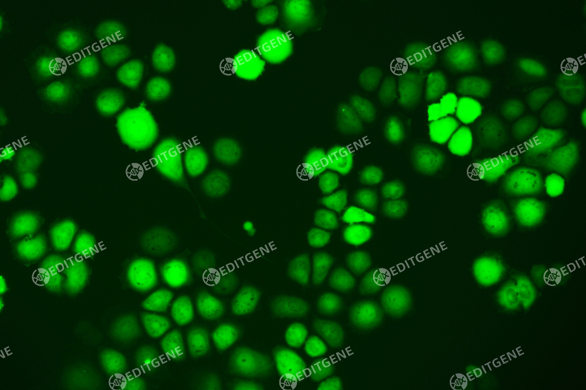
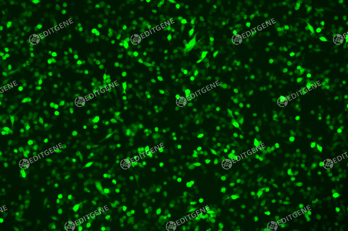
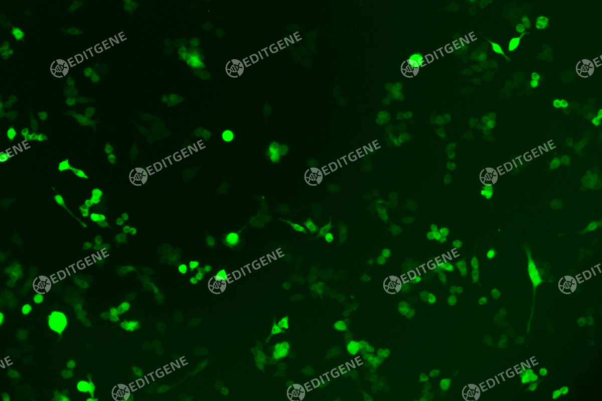
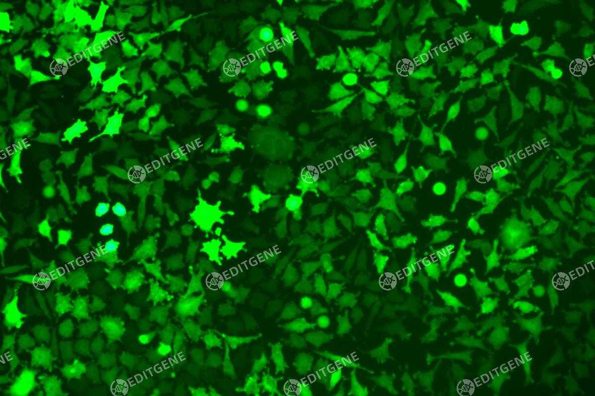
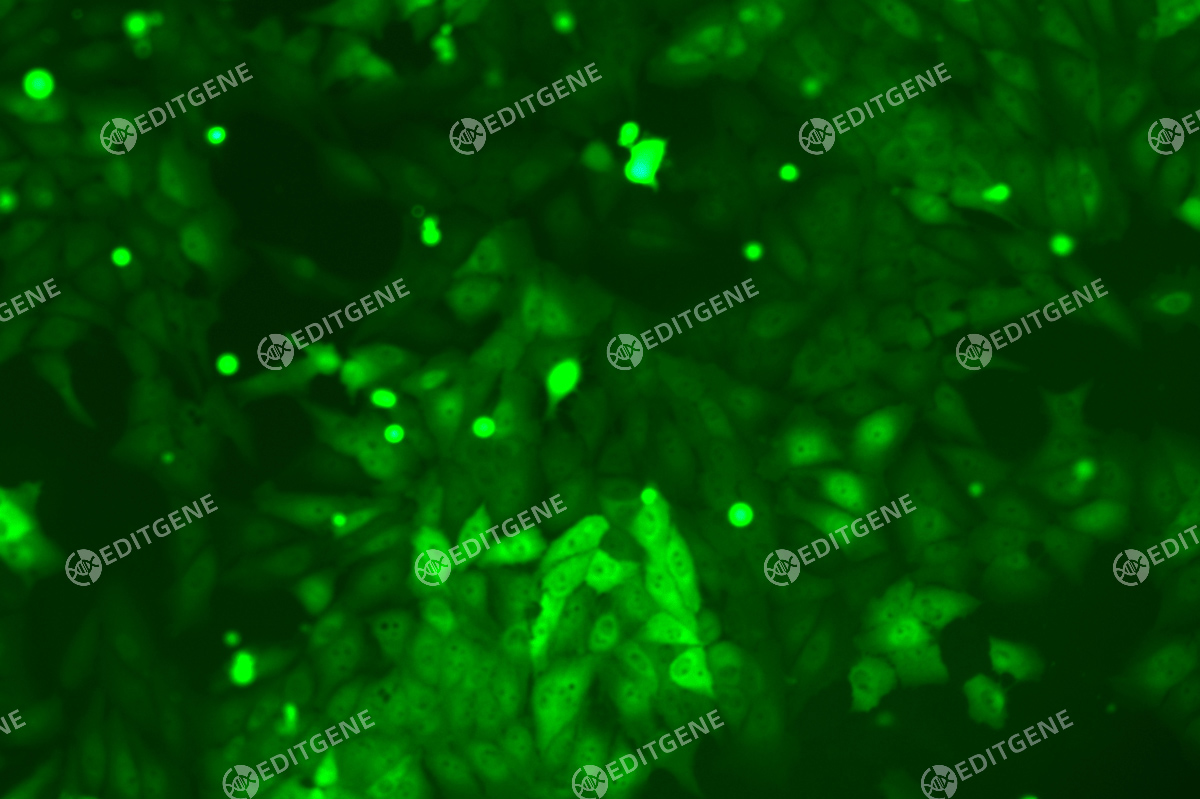
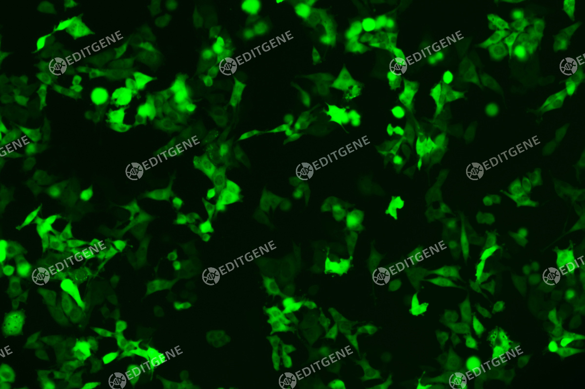
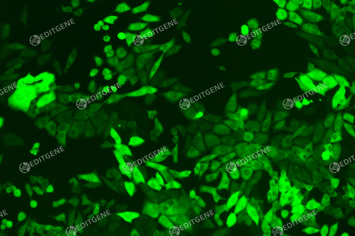
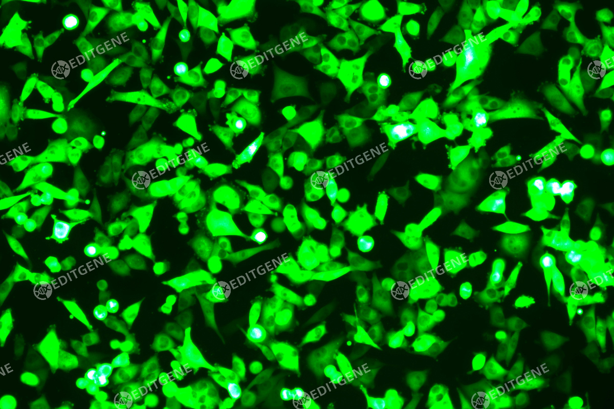
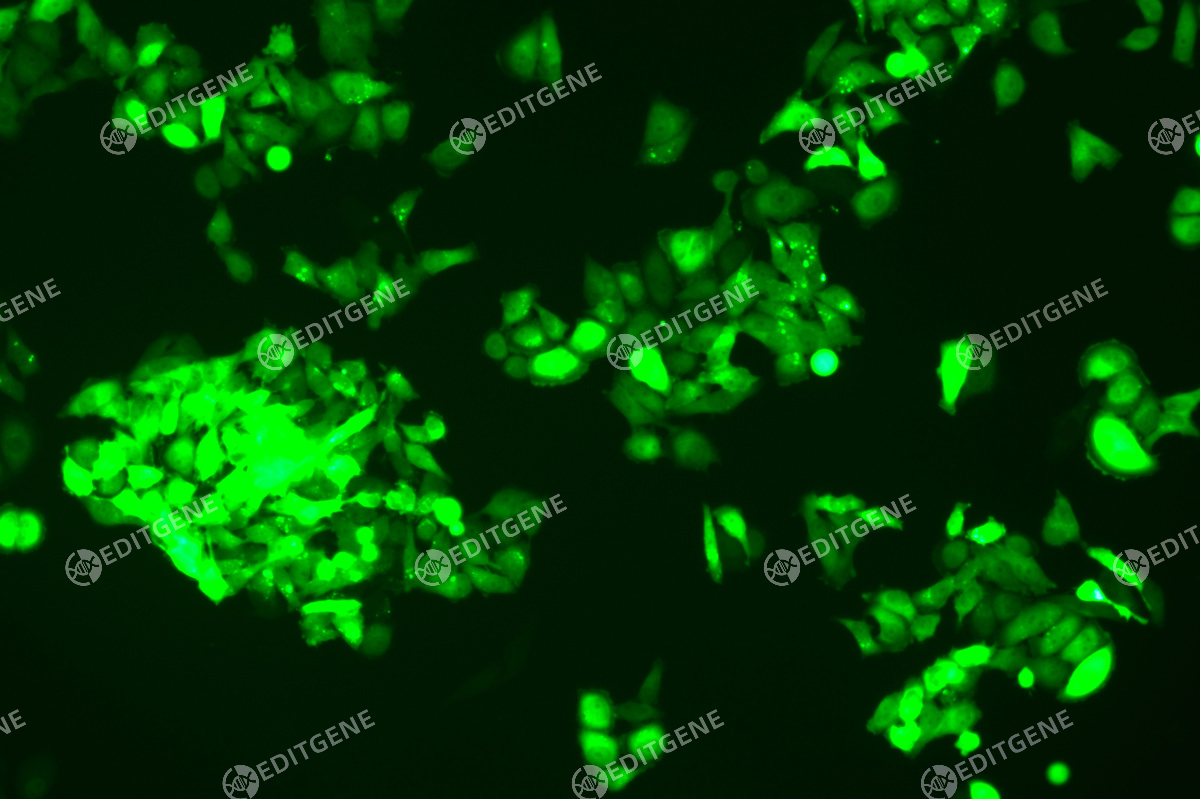
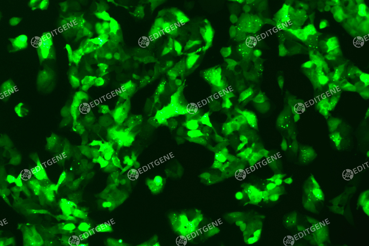
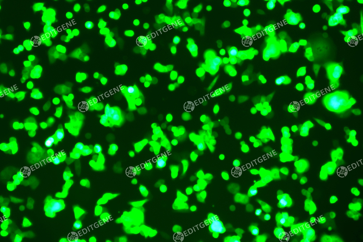
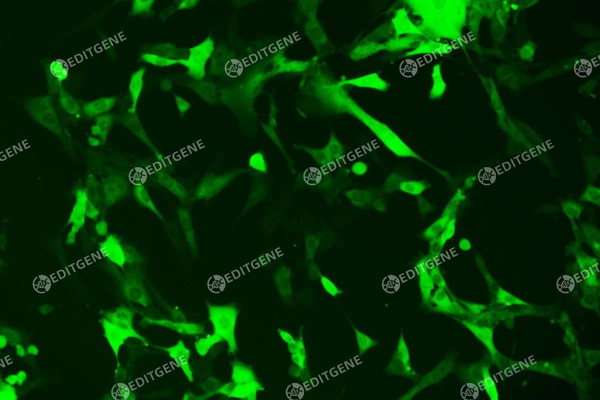
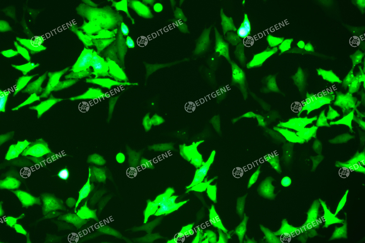
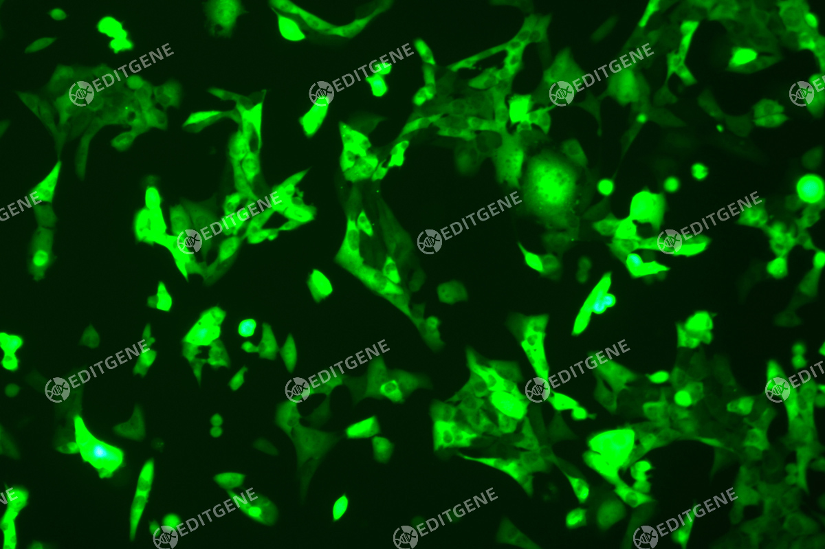
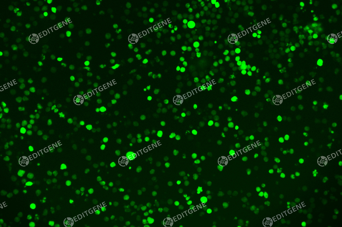
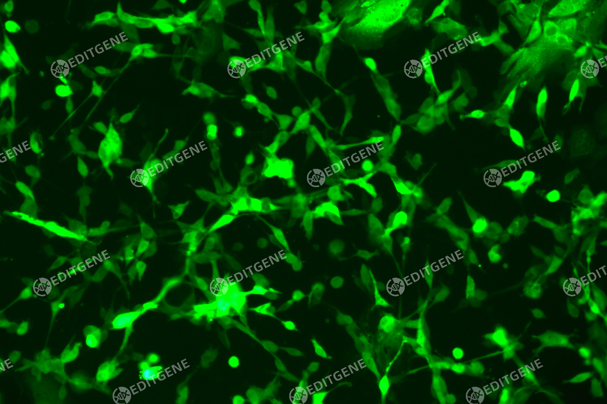
GFP (Green Fluorescent Protein) is a vital tool for observing molecular dynamics within cells. However, it often encounters challenges such as insufficient brightness and limited stability in expression patterns. To tackle these issues, EDITGENE has developed the OE-Booster cis-regulatory element, which enables the production of high-brightness and high-stability CopGFP-tagged cell lines. These cell lines are versatile for use in multiplex fluorescence labeling experiments, enhancing the accuracy of intracellular dynamic observations and improving molecular tracking to reveal complex biological processes.
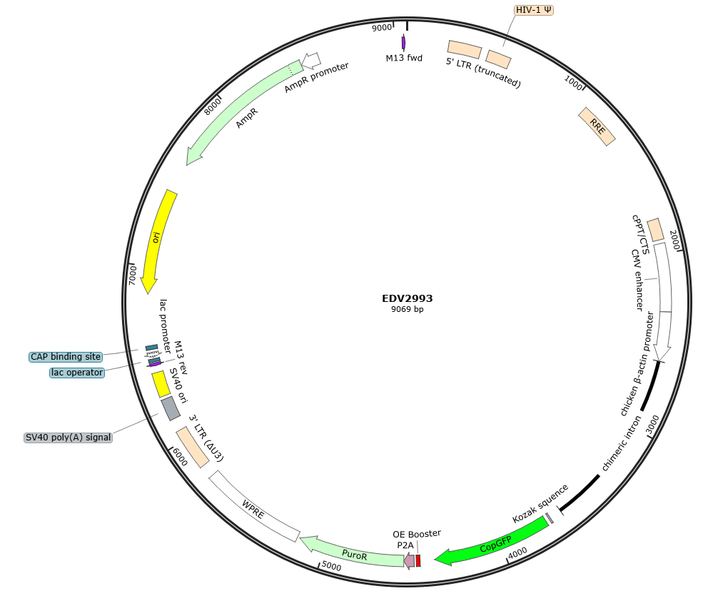
Plasmid Map
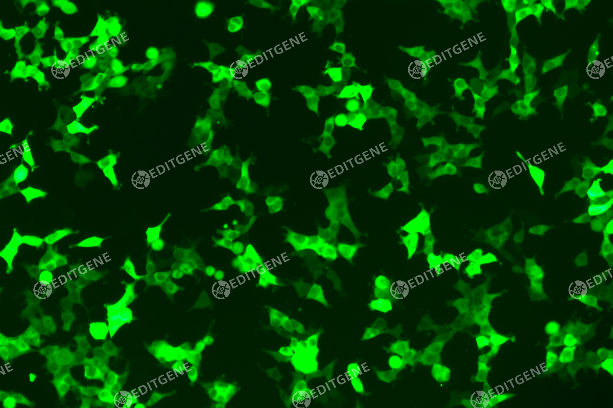
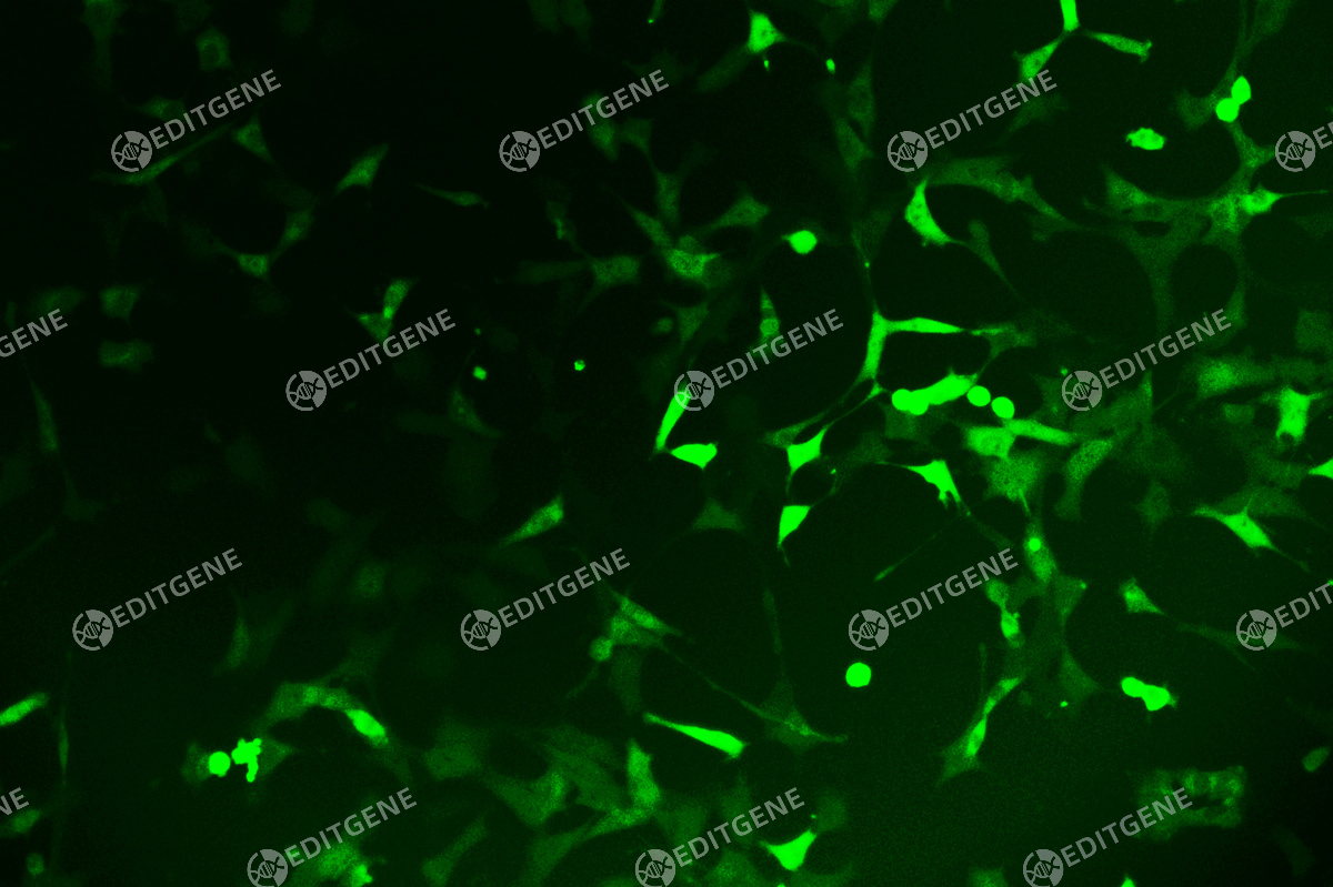
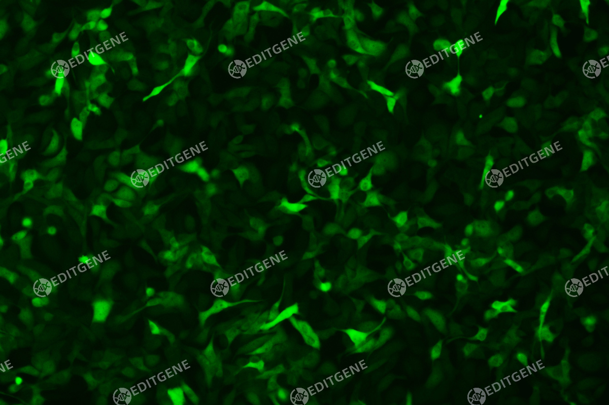
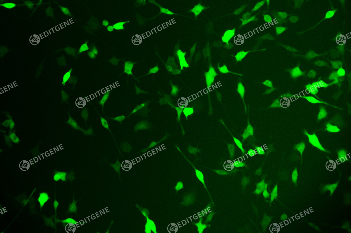
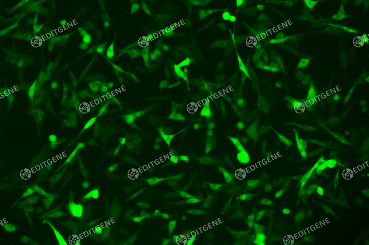
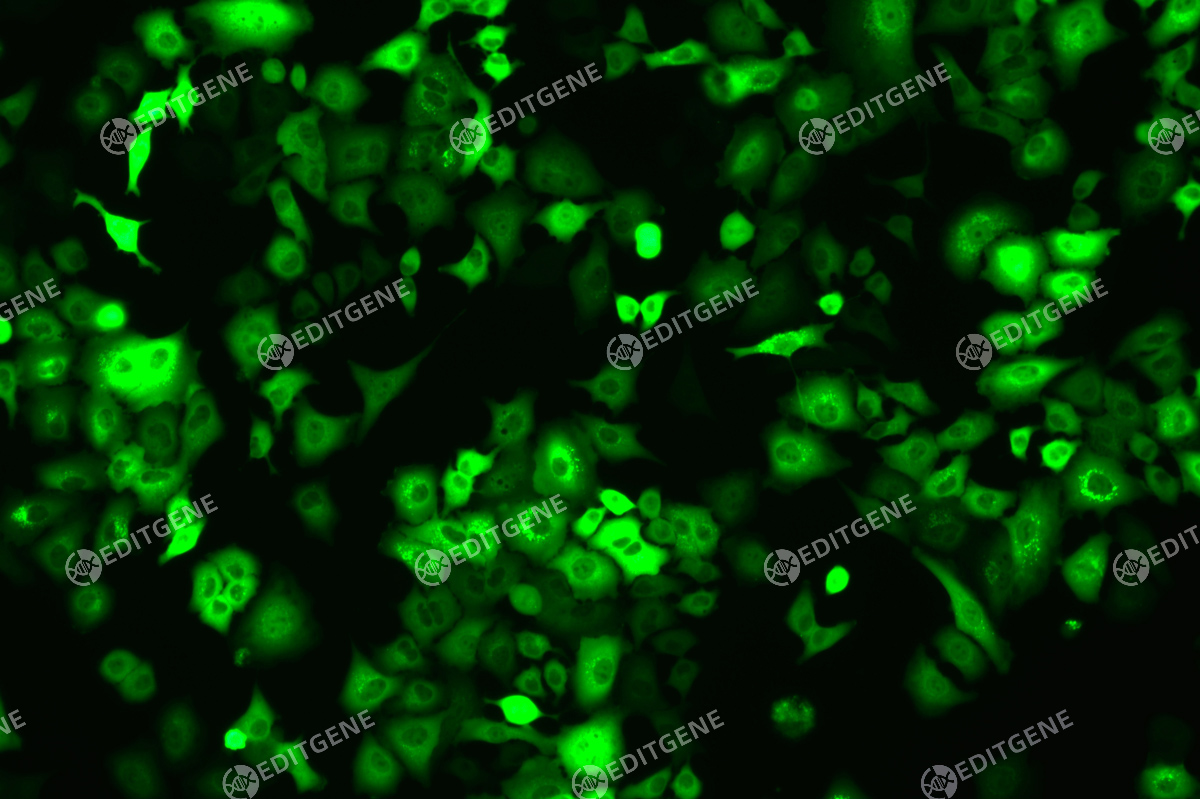
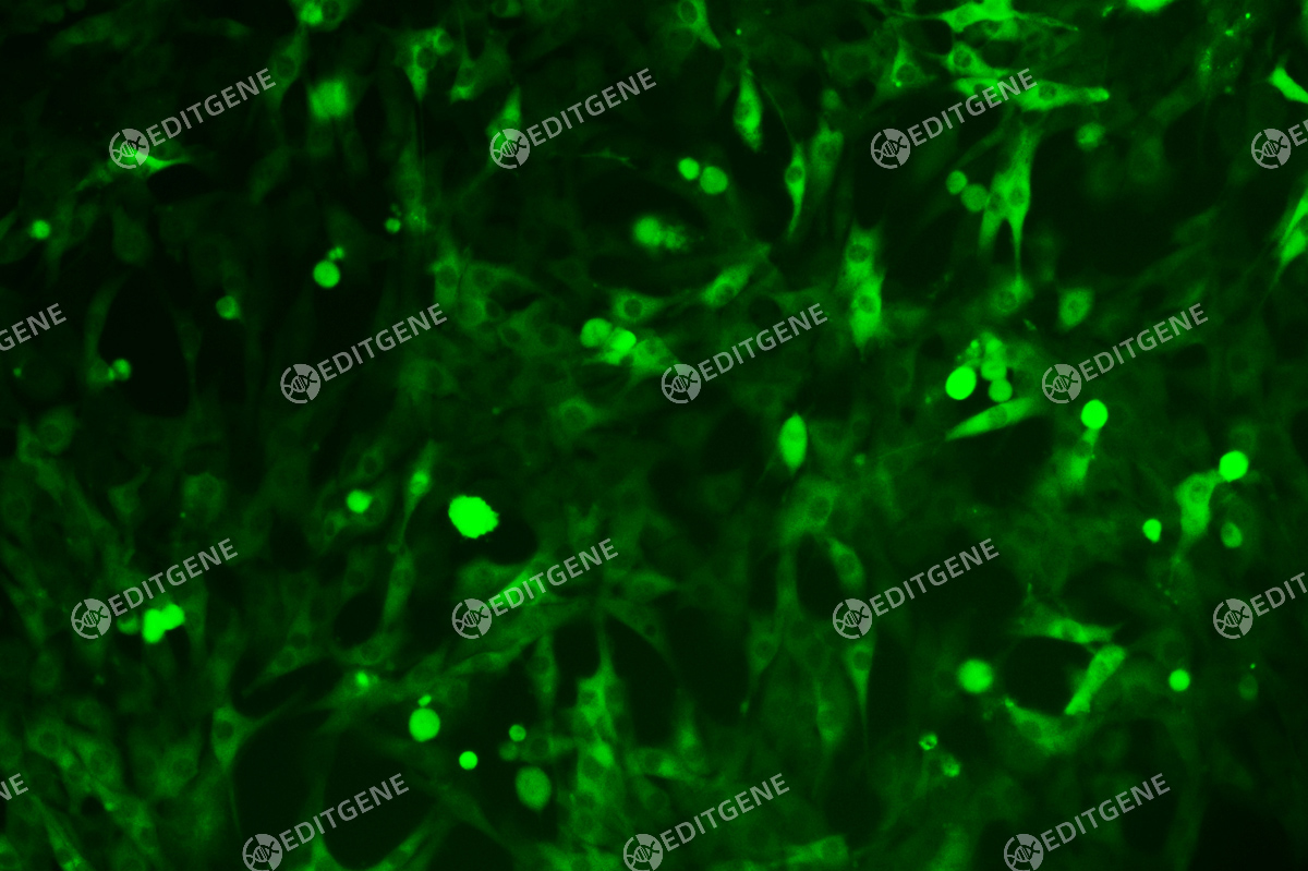
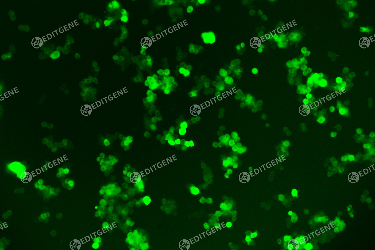
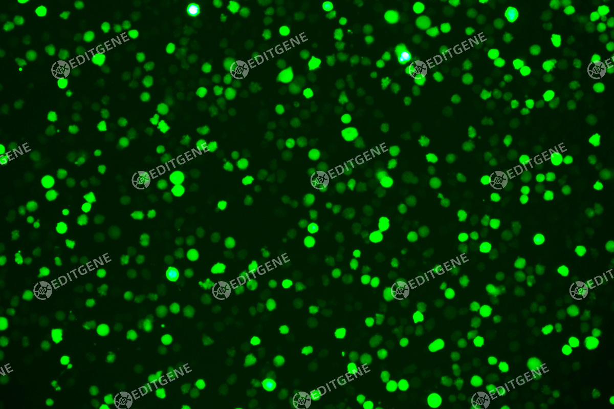
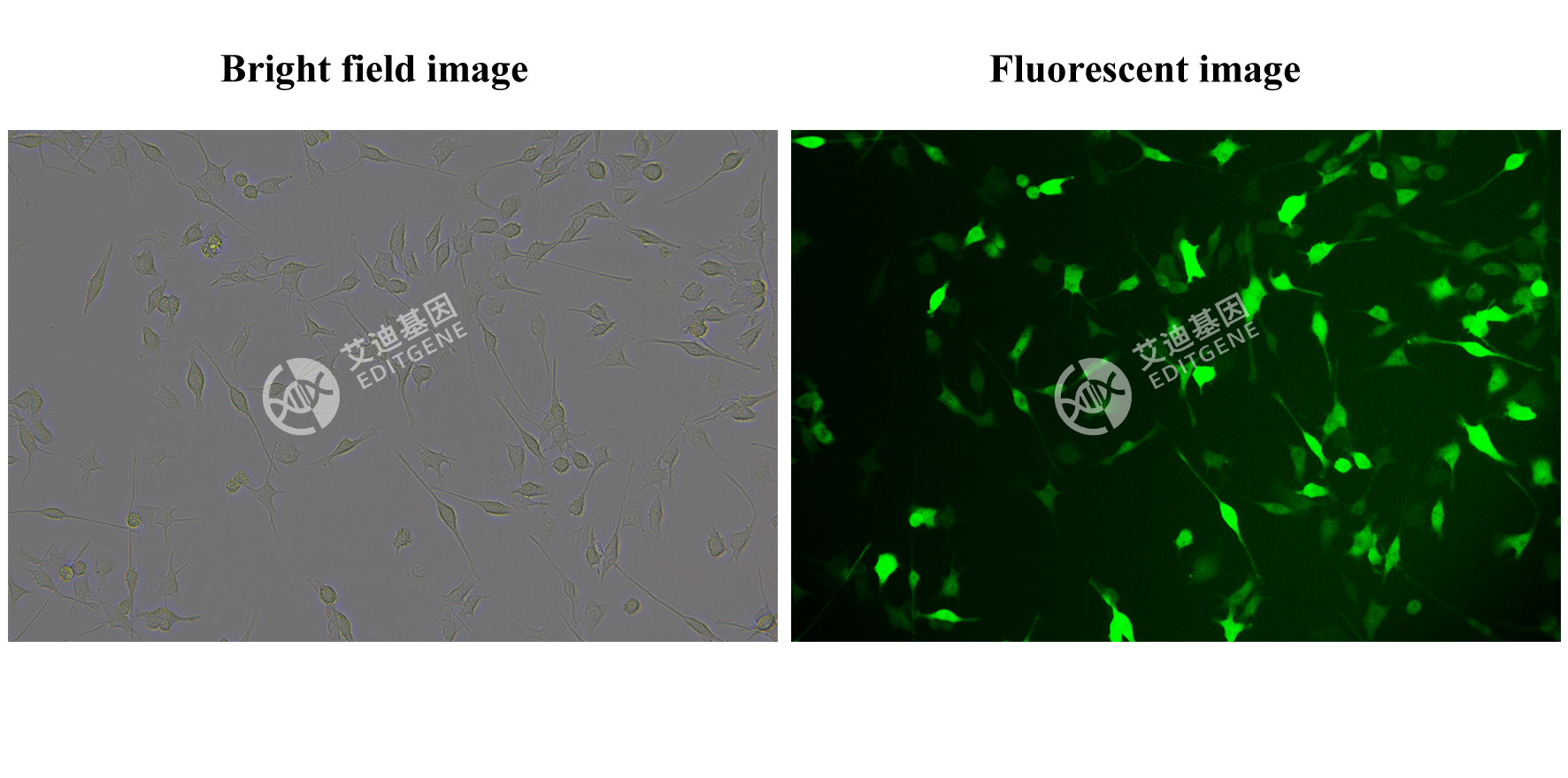
Verification Images
Cell recovery
Note: After receiving the cells, please store in liquid nitrogen. Take it out 10 minutes before recovering cells and place it at -80 ℃ to allow the liquid nitrogen in the tube to evaporate 1. Preheat the water bath and complete culture medium at 37 ℃; 2. Add 6mL of complete culture medium to a 15mL centrifuge tube; 3. Gently swirl the cryopreservation tube in a 37 ℃ water bath until a small piece of ice remains in the cryopreservation tube, please thaw it within 1 minute, and the cap should not touch water; 4. In the ultra clean bench, before opening the cap, wipe the outside of the cryopreservation tube with 75% alcohol; 5. Transfer all the liquid with a pipette to a centrifuge tube of preheated complete culture medium; 6. Rinse the cryopreservation tube with 1mL of complete culture medium to collect residual cells; 7. Centrifuge the cell suspension at 1100 rpm for 4 minutes (centrifugation speed and time depend on cell type); 8. After centrifugation, check if the supernatant is clear and if there is cell pellet at the bottom; inside the hood, carefully pour out the supernatant, add 1mL of complete culture medium, gently resuspend the cell pellet; 9. Evenly seed cells into a T25 flask or culture container with an equivalent bottom area. Add a sufficient amount of complete culture medium, and the total amount of culture medium in a T25 flask shall not be less than 6mL (the actual size of the flask depends on the number of cells frozen in the cryopreservation tube); 10. Gently mix the cells well and place them in a 37 ℃, 5% CO2, saturated humidity incubator (the culture environment depends on cell type and culture medium); 11. Observe the cell status on next day: (1) If the adherent cells adhere well, change fresh complete culture medium; If the cells are observed to be in a round and bright shape but not adhering to the plate, continue cultivation for 24 hours. Afterwards, based on the growth status of the cells, replace the complete culture medium every 2-3 days, observe the cells, and passage cells when confluence rate >80%. If the cell growth is slow or the confluence rate is low, the frequency of medium change can be reduced; (2) When recovering suspension cells, place them in a relatively small container and use a culture medium containing 20% serum for recovery. If the suspended cells are in good condition, change fresh complete culture medium; If the cell status is poor and appears grayscale, it can be further cultured for 72 hours. Change fresh medium if observe live cells. If no significant changes are observed, please contact us for after-sales service in a timely manner.
Passage method
1. View cultures using an inverted phase contrast microscope to assess the degree of confluency and confirm the absence of bacterial and fungal contaminants. Give the flask a gentle knock first, this may dislodge the cells from the flask and remove the need for a trypsinisation step loss of some cells due to associated washes;
2. Decant medium containing non-adherent cells into a sterile centrifuge tube and retain;
3. Wash any remaining attached cells with PBS without Ca2+/Mg2+ using 1-2ml for each 25cm2 of surface area. Retain the washings;
4. Pipette trypsin/EDTA onto the washed cell monolayer using 1ml per 25cm2 of surface area. Rotate flask to cover the monolayer with trypsin. Decant the excess trypsin;
5. Return flask to incubator and leave for 2-10 minutes;
6. Examine the cells using an inverted microscope to ensure that all the cells are detached and floating. The side of the flasks may be gently tapped to release any remaining attached cells;
7. Transfer the cells into the centrifuge tube containing the retained spent medium and cells;
8. Centrifuge the entire cell suspension at 150 x g for 5 minutes;
9. Remove the supernatant and re-suspend the cell pellet in a small volume (10-20ml) of fresh culture medium. Count the cells;
10. Pipette the required number of cells into a new labelled flask and dilute to the required volume using fresh medium;
11. Repeat this process every 2-3 days as necessary.
Storage
Liquid nitrogen


 download

