EDITGENE CO., LTD
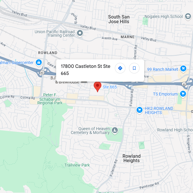
17800 Castleton St. Ste 665. City of Industry. CA 91748
info@editxor.com
+1-833-226-3234 (USA Toll-free)
+1-224-345-1927 (USA)
+86-19120102676 (Intl)

Technical Support

Support Center
Beijing Time: Monday to Friday, 8:00 AM - 6:00 PM
Toll-Free (USA): +833-226-3234
Direct Line (USA): +1-224-345-1927
Email: techsupport@editxor.com

After-Hours Support
Beijing Time: Monday to Sunday, 8:00AM - 6:00 PM
International Line: +86-19120102676
Email: info@editxor.com
Facebook Messenger
Reach out to us on Facebook Messenger for personalized assistance and detailed information.

Linkedin
Engage with us on LinkedIn for professional inquiries, the latest blogs, discoveries, and updates on our innovative work.
FAQ
Which is better for studying SHPRH function, SHPRH Knockout HAP1 Cell Line or SHPRH overexpression HAP1 Cell Line?
The choice depends on whether you are studying SHPRH's role as a ubiquitin ligase mediating PCNA polyubiquitination during DNA damage tolerance or its functions in genome stability maintenance. The Knockout line is the standard tool for asking whether SHPRH is required for K63-linked polyubiquitination of PCNA at lysine 164 — SHPRH and HLTF redundantly catalyze this modification, which switches translesion synthesis from error-prone to template-switching error-free pathway. Overexpression is useful for testing SHPRH activity or for studying its tumor suppressor functions.
For DNA damage tolerance research, the EDITGENE SHPRH Knockout in HAP1 is a clean genetic background for studying PCNA-K164 polyubiquitination biology. HLTF expression analysis is essential given functional redundancy — combined SHPRH/HLTF analyses provide comprehensive characterization of K63-polyubiquitin-dependent template switching. Rescue with wild-type or RING-domain-mutant (E3-ligase-dead) SHPRH enables structure-function studies.
What are the application scenarios for this model?
Primary applications:
• PCNA polyubiquitination: ubiquitin Western blot for K63-polyubiquitinated PCNA following DNA damage (UV, methyl methanesulfonate) — assess SHPRH-dependent modification.
• Template switching activity: replication fork progression assays following damage induction to assess damage tolerance pathway integrity.
• HLTF paralog studies: HLTF expression analysis and combined SHPRH/HLTF knockdown to dissect redundant K63-polyubiquitination roles.
• Genome stability: micronucleus assays, sister chromatid exchange, and damage-induced mutagenesis given SHPRH's reported tumor suppressor functions.
EDITGENE recommends this model for researchers investigating DNA damage tolerance, PCNA polyubiquitination, and SHPRH-mediated genome stability.
Is this SHPRH Knockout HAP1 Cell Line compatible with overexpression rescue experiments?
Yes. SHPRH rescue experiments require attention to multi-domain architecture:
• Construct design: SHPRH is large (~1683 amino acids); use codon-modified sequences with C-terminal tags (FLAG, HA). The N-terminal H15 (linker histone H1.5) domain, central SF2 helicase domain, RING domain (E3 ligase), and C-terminal regions should all be preserved.
• RING-dead rescue: RING domain mutations abolish E3 ligase activity and are the standard specificity control for K63-polyubiquitin ligase function.
• Helicase-dead rescue: ATP-binding mutations in the SF2 domain enable separation of ATPase from E3 ligase functions.
• Functional readout: rescue should restore K63-polyubiquitinated PCNA following DNA damage and template switching activity.
HAP1-specific considerations:
• Diploidization: HAP1 cells gradually diploidize during extended culture — confirm ploidy by flow cytometry at the time of phenotypic assay.
• Integration site sensitivity: position effects on transgene expression are more pronounced in near-haploid backgrounds; generating multiple independent rescue clones is strongly recommended.
• Transduction efficiency: HAP1 transduces with lentivirus at moderate efficiency — increase MOI compared to standard immortalized lines.
Which is better for studying SCNN1D function, SCNN1D Knockout HAP1 Cell Line or SCNN1D overexpression HAP1 Cell Line?
The choice depends on whether you are studying SCNN1D (ENaC δ subunit)'s role in heteromeric epithelial sodium channel assembly or its specific contributions as a tissue-restricted ENaC subunit. The Knockout line is appropriate for asking whether SCNN1D is required for δ-containing ENaC channel activity — δ-ENaC is expressed primarily in apical membranes of pancreas, brain, ovary, and testis, with proton-activated and capsaicin-sensitive properties distinct from canonical α-ENaC channels. Overexpression is useful for testing channel activity in heterologous systems or for assembly studies with β-ENaC and γ-ENaC.
For ENaC research, the EDITGENE SCNN1D Knockout in HAP1 enables mechanistic studies of δ-ENaC biology — δ-ENaC differs from the more widely studied α-ENaC (SCNN1A) and has been implicated in proton sensing and pain modulation. Rescue with wild-type or pore-mutant SCNN1D, with β/γ-ENaC co-expression, enables comprehensive channel assembly and function studies.
What are the application scenarios for this model?
Primary applications:
• Sodium current assays: patch-clamp electrophysiology to measure δ-containing ENaC currents — δ-ENaC channels have distinct sensitivity to amiloride and proton activation compared to α-ENaC.
• Heterologous ENaC reconstitution: combined SCNN1D + SCNN1B + SCNN1G expression to characterize δβγ-ENaC channel properties.
• Proton-sensitive transport: pH-dependent sodium current studies given δ-ENaC's reported proton activation.
• Tissue-restricted ENaC biology: heterologous studies in HAP1 background as a clean genetic context.
EDITGENE recommends this model for researchers investigating δ-ENaC biology and tissue-specific epithelial sodium channel function.
Is this SCNN1D Knockout HAP1 Cell Line compatible with overexpression rescue experiments?
Yes. SCNN1D rescue experiments require attention to ENaC subunit assembly:
• Construct design: use a codon-modified SCNN1D sequence with a small intracellular tag (FLAG, HA). δ-ENaC is a type II membrane protein with two transmembrane domains and large extracellular loop — preserve all elements.
• Subunit co-expression: δ-ENaC requires β-ENaC (SCNN1B) and γ-ENaC (SCNN1G) for functional channel formation — rescue lines should be characterized for β/γ subunit expression, or co-rescue may be needed.
• Pore-mutant rescue: selectivity filter mutations enable distinguishing channel function from non-conducting roles.
• Functional readout: rescue should restore amiloride-sensitive sodium currents and proton-activated channel function in heterologous reconstitution.
HAP1-specific considerations:
• Diploidization: HAP1 cells gradually diploidize during extended culture — confirm ploidy by flow cytometry at the time of phenotypic assay.
• Integration site sensitivity: position effects on transgene expression are more pronounced in near-haploid backgrounds; generating multiple independent rescue clones is strongly recommended.
• Transduction efficiency: HAP1 transduces with lentivirus at moderate efficiency — increase MOI compared to standard immortalized lines.
Which is better for studying RXRA & RXRB function, RXRA & RXRB Knockout A-549 Cell Line or RXRA & RXRB overexpression A-549 Cell Line?
The choice depends on whether you are studying combined RXR-mediated nuclear receptor signaling in lung cancer biology or distinguishing RXR functions from RXRG-independent processes. The Double Knockout line is uniquely valuable for asking whether RXR-mediated transcription is required for these processes — combined RXRA + RXRB loss eliminates the dominant RXR partners in most non-hematopoietic cells, leaving only minimal RXRG (typically tissue-restricted) compensation. Overexpression of either RXR isoform in the double knockout enables isoform-specific functional dissection.
For lung cancer nuclear receptor research, the EDITGENE RXRA & RXRB Double Knockout in A-549 is highly informative — A-549 is a workhorse NSCLC model, and the double knockout disrupts retinoid X receptor partnerships with RAR (retinoid signaling), PPAR (lipid metabolism), LXR (cholesterol homeostasis), and other partners simultaneously. Single-RXR rescue (RXRA alone or RXRB alone) in the double knockout is the gold-standard experimental design for distinguishing paralog-specific functions. The knockout is valuable for studying bexarotene (Targretin), RXR-selective rexinoid mechanism, and emerging RXR-targeted therapeutics.
What are the application scenarios for this model?
Primary applications:
• Combined retinoid/rexinoid response: RAR-RXR (retinoic acid response) and RXR-RXR (rexinoid response) reporter assays simultaneously disrupted in the double knockout.
• Single-RXR rescue studies: re-introduction of RXRA alone or RXRB alone enables isoform-specific functional dissection in a clean double-knockout background — the gold-standard experimental design.
• Nuclear receptor partner studies: PPAR, LXR, FXR, VDR, TR signaling readouts to assess pan-RXR partnership disruption.
• Bexarotene/rexinoid specificity: critical genetic control for RXR-targeted compounds (bexarotene, IRX4204, A-792611) in cancer and metabolic disease contexts.
• Lung cancer biology: differentiation, proliferation, and EMT studies given RAR-RXR signaling's role in lung cancer biology.
EDITGENE recommends this model for researchers investigating RXR family biology, RXR-targeted nuclear receptor pharmacology, and lung cancer-relevant retinoid signaling.
Is this RXRA & RXRB Knockout A-549 Cell Line compatible with overexpression rescue experiments?
Yes, and rescue experiments are uniquely powerful in this double knockout background:
• Single-RXR rescue: re-introduction of RXRA alone or RXRB alone in the double knockout enables isoform-specific functional dissection — the gold-standard experimental design for redundant nuclear receptor paralogs.
• Construct design: codon-modified RXRA or RXRB sequences with small C-terminal tags (FLAG, HA). Preserve all nuclear receptor functional domains.
• DNA-binding-deficient and ligand-binding-deficient rescue: structure-function variants enable dissection of RXR-specific activities in the clean double-knockout background.
• Functional readout: rescue should restore RAR-RXR retinoid signaling (DR5 reporter), PPAR-RXR lipid metabolism signaling (DR1 reporter), and other partnered nuclear receptor activities.
A-549 transduces efficiently with lentivirus and supports systematic single-paralog rescue experiments that would be difficult to interpret in single-knockout backgrounds with paralog compensation.
Which is better for studying SLC16A12 function, SLC16A12 Knockout Huh-7 Cell Line or SLC16A12 overexpression Huh-7 Cell Line?
The choice depends on whether you are studying SLC16A12 (MCT12)'s role as a creatine transporter or modeling autosomal dominant juvenile cataracts. The Knockout line is the standard tool for asking whether MCT12 is required for creatine transport — MCT12 has been identified as a creatine transporter distinct from the better-known CRT1/SLC6A8. Overexpression is useful for testing transport activity or for studying disease-associated mutations.
For creatine biology research, the EDITGENE MCT12 Knockout in Huh-7 enables study of MCT12-mediated creatine handling. SLC16A12 mutations cause autosomal dominant juvenile cataracts, glucosuria, and elevated guanidinoacetate — disease variant rescue enables genotype-function correlation. SLC6A8 (CRT1) expression analysis aids interpretation given functional overlap in creatine transport.
What are the application scenarios for this model?
Primary applications:
• Creatine uptake assays: ³H-creatine uptake measurement to quantify MCT12 transport activity.
• Disease modeling: rescue with autosomal dominant juvenile cataract-associated SLC16A12 mutations for genotype-function studies.
• Substrate scope characterization: testing related substrates given MCT12's emerging substrate profile.
• Paralog studies: SLC6A8 (CRT1) expression analysis given the two transporters' overlap in creatine handling.
EDITGENE recommends this model for researchers investigating creatine transport biology and SLC16A12-related cataract disease mechanisms.
Is this SLC16A12 Knockout Huh-7 Cell Line compatible with overexpression rescue experiments?
Yes. MCT12 rescue experiments require attention to creatine transport biology:
• Construct design: use a codon-modified SLC16A12 sequence with a small C-terminal tag (FLAG, HA). The 12-transmembrane SLC16 architecture must be preserved.
• Disease mutation rescue: cataract-associated SLC16A12 mutations enable genotype-function correlation studies.
• Transport-deficient rescue: substrate-binding pocket mutations enable structure-function studies.
• Functional readout: rescue should restore creatine uptake activity measured by ³H-creatine uptake.
Huh-7 transduces efficiently with lentivirus and supports stable rescue line generation.
Which is better for studying SIRT7 function, SIRT7 Knockout HAP1 Cell Line or SIRT7 overexpression HAP1 Cell Line?
The choice depends on whether you are studying SIRT7's role as a nucleolar NAD⁺-dependent deacetylase regulating ribosomal RNA transcription or its emerging functions in DNA damage response, metabolism, and aging biology. The Knockout line is the standard tool for asking whether SIRT7 is required for H3K18ac deacetylation, RNA polymerase I transcription regulation, or its other reported functions. Overexpression is useful for studying SIRT7 in cancer contexts where it has been reported to be upregulated.
For sirtuin research, the EDITGENE SIRT7 Knockout in HAP1 provides a clean genetic background for dissecting SIRT7-specific functions among the seven mammalian sirtuins. Rescue with wild-type or catalytically-dead (H187Y) SIRT7 is the standard specificity control. SIRT7 has emerging interest in aging, NAD⁺ biology, and cancer — the knockout serves as a critical specificity tool for SIRT7-selective compounds in development.
What are the application scenarios for this model?
Primary applications:
• H3K18ac deacetylation: histone H3K18 acetylation Western blot/ChIP analysis at SIRT7 target gene promoters.
• Ribosomal RNA transcription: 47S pre-rRNA quantification by qPCR to assess SIRT7's effect on Pol I transcription.
• NAD⁺-dependent activity: in vitro deacetylation assays with NAD⁺ supplementation and varied conditions.
• Cancer biology: proliferation, transformation, and stress response phenotypes given SIRT7's reported tumor-supportive functions.
EDITGENE recommends this model for researchers investigating sirtuin biology, nucleolar function, NAD⁺-dependent deacetylase mechanisms, and SIRT7-targeted compound development.
Is this SIRT7 Knockout HAP1 Cell Line compatible with overexpression rescue experiments?
Yes. SIRT7 rescue experiments are well-established for sirtuin research:
• Construct design: use a codon-modified SIRT7 sequence with a small C-terminal tag (FLAG, HA). SIRT7 has a central catalytic domain with N- and C-terminal extensions critical for nucleolar localization and substrate recognition.
• Catalytically-dead rescue: the H187Y mutation abolishes NAD⁺-dependent deacetylase activity and is the standard specificity control.
• Nucleolar localization validation: confirm nucleolar localization by fibrillarin co-staining before functional assays.
• Functional readout: rescue should restore H3K18ac levels at SIRT7 target promoters and ribosomal RNA transcription.
HAP1-specific considerations:
• Diploidization: HAP1 cells gradually diploidize during extended culture — confirm ploidy by flow cytometry at the time of phenotypic assay.
• Integration site sensitivity: position effects on transgene expression are more pronounced in near-haploid backgrounds; generating multiple independent rescue clones is strongly recommended.
• Transduction efficiency: HAP1 transduces with lentivirus at moderate efficiency — increase MOI compared to standard immortalized lines.

Room 501, Building D, International Business Incubator, No.3 Juquan Road, Science City, Huangpu District, Guangzhou, Guangdong, China 510663